
 772-228-6882




Your knee’s movement depends on several structures working together. Understanding knee bone anatomy gives you a practical framework for making sense of discomfort. The bones provide structure, while cartilage and surrounding soft tissues support movement. No single sensation tells the whole story, but knowing the basic layout can help you describe where symptoms occur and which activities bring them on.
You may notice discomfort while walking, bending, or getting up from a chair and wonder which part of the knee is involved. Anatomy offers context, not a diagnosis. The location and pattern of symptoms, together with a clinical assessment, help distinguish possible sources and support a more informed conversation about your mobility.
This guide explains how the knee’s key structures relate to one another and why they matter in everyday movement. You’ll build a practical map of the joint, learn how muscles contribute to motion, and gain clearer language for discussing persistent symptoms and possible next steps with a qualified healthcare professional.
Key Takeaways
- Learn how smooth joint surfaces help the knee move with less friction.
- See how the menisci cushion the joint and spread forces across a wider area.
- Understand why the fibula is near the knee but isn’t part of its main hinge joint.
- Trace how opposing muscle groups coordinate to control bending and straightening.
Articular cartilage
Articular cartilage covers the bone surfaces that meet in the knee, creating a smooth interface as the joint moves. Its location and condition help explain how the joint handles motion and load.
Femoral Condyles and Joint Surfaces
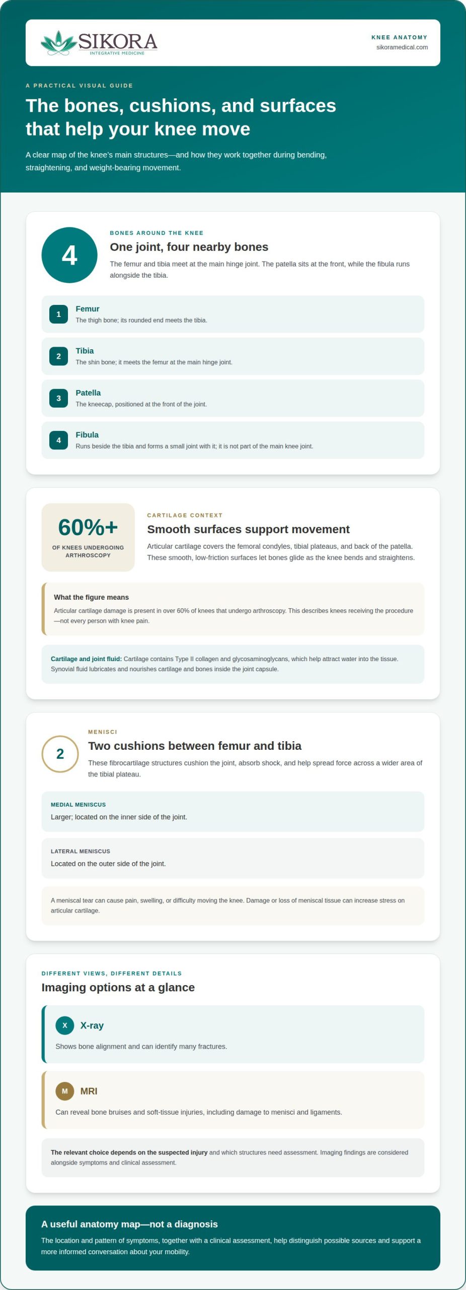
The femur ends in two rounded condyles that meet the tibial plateau. Articular cartilage covers these condyles, the tibial plateaus, and the back of the patella. These coated surfaces let bones glide as the knee bends and straightens. The tibial plateau has medial and lateral regions, corresponding to the inner and outer sides of the joint. This map helps describe where a cartilage finding appears, although location alone does not establish the cause of pain.
Cartilage Function in Bone Articulations
Articular cartilage provides a smooth, low-friction covering where bones articulate. It contains Type II collagen and glycosaminoglycans, molecules that help attract water into the tissue. That water-rich structure helps cartilage respond to pressure during movement. The knee also has fibrocartilage in its menisci, distinct from the hyaline articular cartilage covering bone surfaces. Synovial fluid, produced by the joint’s synovial membrane, lubricates and nourishes cartilage and bones inside the joint capsule. Together, these features support the moving joint.
Cartilage and Arthritis
Arthritis can involve damage or wear to articular cartilage, changing the surfaces that move against each other. Cartilage has no blood vessels and a very low capacity for healing, so damage may persist rather than repair like tissue with a richer blood supply. Articular cartilage damage is present in over 60% of knees that undergo arthroscopy. That figure describes knees receiving the procedure, not every person with knee pain. Symptoms and clinical findings help put cartilage changes in context.
Knee Joint Anatomy: What Is a Knee & What Does It Do?
Common knee bone injuries include fractures and bone bruises, which affect bone differently. X-rays can show many fractures; MRI can reveal bone bruises and soft-tissue injuries, including meniscus damage.
Meniscus Structure and Cushioning
The menisci are fibrocartilage cushions located between the femur and tibia. They cushion the joint during activities such as walking, running, and jumping by absorbing shock. Their shape also helps the knee stay steady as it bends and straightens. Because the menisci sit between the bones, damage can affect comfort and movement as well as cushioning.
Menisci and Load Distribution
Each meniscus spreads force across a wider area of the tibial plateau instead of concentrating pressure in one small spot. This load distribution helps protect the joint surfaces during weight-bearing movement. The medial meniscus is larger than the lateral meniscus, and both work alongside other structures. The posterior cruciate ligament, for example, helps control movement between the femur and tibia, while the fibula lies alongside the tibia.
A meniscal tear can cause pain, swelling, or difficulty moving the knee. Damage to or loss of meniscal tissue can increase stress on articular cartilage and may contribute to cartilage breakdown and arthritis. Surgery is only one consideration: the injury, symptoms, and effects on daily movement all matter when assessing treatment options.
Imaging Bone Anatomy and Injuries
X-rays provide images of bone alignment and can identify many fractures, while MRI can show bone bruises, menisci, ligaments, and other soft tissues. These tests offer different views, so the relevant choice depends on the suspected injury and the structures that need assessment. For example, after a twisting injury, imaging may help distinguish a bone injury from damage to the medial meniscus or a ligament. If knee symptoms persist, pain management care can be a next step in discussing evaluation and treatment options.
Bones of the knee
Understanding knee bone anatomy starts with the main joint and the nearby bones that support it. The femur and tibia meet at the knee, with the patella at the front and the fibula alongside. During childhood growth, growth plates allow bones to lengthen; throughout life, bone tissue continues to remodel.
The Four Knee Bones
The four bones around the knee are the femur, tibia, patella, and fibula. The femur meets the tibia at the main hinge joint, while the patella sits in a groove at the front of the femur. The fibula runs beside the tibia and forms a small joint with it, but it is not part of the main knee joint. Knowing which bone is involved makes anatomy and injury descriptions clearer.
Patella Movement and Protection
The patella, or kneecap, sits within the quadriceps tendon and moves through the femur’s trochlear groove as your knee bends and straightens. It protects the front of the joint and acts as a fulcrum, improving the quadriceps’ pull when they extend your knee. The quadriceps tendon attaches above the patella, while the patellar tendon connects it to the tibia. This arrangement supports controlled movement, such as standing up.
Bone Development and Age-Related Changes
In children, growth plates near the ends of the femur and tibia allow those bones to lengthen. As growth ends, the plates become solid bone, while bone tissue continues to remodel in response to the body’s needs. With age, changes in bone and joint surfaces may affect how the knee feels or moves. These developments are part of knee bone anatomy, but a symptom’s location alone cannot identify the structure involved.
Bones rely on ligaments and tendons for support and movement. Ligaments connect bone to bone; the medial collateral ligament, for example, helps support the inner knee. Tendons connect muscle to bone, transferring force to move the joint. The fibula also provides attachment sites for the lateral collateral ligament and biceps femoris tendon. These relationships add another layer to knee bone anatomy: stability and movement involve more than bones alone.
Knee flexor and extensor muscles
Your knee’s muscles create and control its main movements. The quadriceps at the front of the thigh are extensors because their contraction straightens the knee. The hamstrings at the back are flexors because their contraction bends it. Their opposing actions let you move between bent and straight positions with control. Tendons connect these muscles to bone and transfer their pulling force.
The quadriceps consist of four muscles that work together to extend the knee. Their force travels through the quadriceps tendon to the patella, then through the patellar tendon to the tibia. The kneecap acts as a lever, helping the quadriceps extend the leg. When you rise from a chair, for example, the quadriceps contract and pull through this tendon chain to straighten your knee. If the movement feels uncomfortable, the muscles are only one part of the picture.
The hamstrings consist of three muscles at the back of the thigh. Their tendons attach around the knee, and the muscles contract to bend it. You use this action to bring your heel toward your body or draw your lower leg back. In everyday movement, the hamstrings and quadriceps coordinate: one group produces the main action while the other helps control the transition. Together, they support bending and straightening without reducing the knee to a simple hinge.
Muscle action also occurs alongside the knee’s ligaments, including the ACL and PCL, which connect the femur and tibia and help control their relative movement. The patella, quadriceps tendon, and patellar tendon form a force pathway, while the hamstrings provide a countering pull from behind. This anatomy helps you describe whether discomfort appears during bending, straightening, or both, but the activity alone cannot identify its cause. If knee symptoms are limiting your movement, discuss your knee pain and movement concerns with a medical professional.
Sikora Integrative Medicine: Stem Cells
Sikora Integrative Medicine offers Stem Cells and Exosomes as regenerative medicine options for people exploring care for musculoskeletal concerns. If knee symptoms continue to affect your daily movement, a clinical discussion can connect your symptoms and evaluation findings with possible next steps. The team discusses evidence and regulatory status so you can consider what an option involves and how it relates to your goals.
Not every knee concern calls for the same approach; the source of symptoms and your overall health matter. Stem Cells or Exosomes warrant an informed discussion, not an assumption that they will resolve the problem or replace understanding its cause. Contact Sikora Integrative Medicine to discuss your knee concerns and care options.
Take the Next Step Toward More Comfortable Movement
A clearer understanding of knee bone anatomy can help you describe what you feel, but persistent discomfort calls for more than anatomy alone. The next step is to consider your symptoms, movement limits, and clinical findings together. This discussion can clarify whether an option suits your situation, rather than assuming one approach fits every knee concern.
At Sikora Integrative Medicine, Dr. Alita Sikora’s credentials and treatment areas provide context for the team’s care. Dr. Alita Sikora’s team offers Stem Cells and PRP as part of its regenerative medicine services. Hormone Therapy is also available for patients addressing hormone-related concerns. These options serve different purposes, so discussing your health goals and evaluation findings can help guide your choices.
If knee symptoms are affecting your daily life, take a measured next step and discuss your concerns with the team. Contact Sikora Integrative Medicine to start the conversation, and move forward with a clearer understanding of your options.
Frequently Asked Questions
Which bones make up the knee joint?
The femur, tibia, and patella form the main knee joint, while the fibula runs alongside it. The fibula meets the tibia at a small joint but is not part of the main hinge. This distinction can help you understand an imaging report: a finding near the fibular head may involve a neighboring bone rather than the knee’s main weight-bearing surfaces.
What are the parts of the femur, tibia, and patella at the knee?
At the knee, the femur ends in rounded condyles, the tibia has a broad upper plateau, and the patella sits at the front. The femoral condyles meet the tibial plateau, while the kneecap glides against the femur. These landmarks help describe the location of a symptom or imaging finding, such as the front of the knee versus its inner or outer side.
How do the femur and tibia meet to form the knee joint?
The lower end of the femur meets the upper end of the tibia to form the tibiofemoral articulation. This connection lets your knee bend and straighten, with slight rotation when bent. The patella also meets the femur at a separate patellofemoral articulation. These contact points help explain why discomfort during a squat may feel different from pain at the front of the kneecap.
What are the medial and lateral femoral condyles and tibial plateaus?
The medial condyle and plateau are on the inner side of the knee; the lateral pair are on the outer side. Each femoral condyle meets its corresponding tibial plateau. These paired regions help clinicians describe the location of tenderness or an imaging finding precisely. For example, “medial” means the side nearer your other knee, not a separate bone.
How do the menisci fit between the knee bones?
The medial and lateral menisci sit between the femoral condyles and tibial plateau, along the inner and outer sides of the joint. Their crescent shapes do not cover the entire tibial surface. They help distribute forces and support joint stability alongside the ligaments. A twist or deep bend can stress a meniscus, but symptoms alone cannot confirm a tear.
What ligaments connect and stabilize the bones of the knee?
The knee’s four major ligaments are the anterior cruciate ligament, posterior cruciate ligament, medial collateral ligament, and lateral collateral ligament. The cruciate ligaments cross within the joint and help control forward and backward movement between the tibia and femur. The collateral ligaments run along the inner and outer sides, limiting excessive side-to-side movement. Together, they help stabilize the knee as you change direction.

Article by
Alita G. Sikora, MD
Dr. Alita Gonsalves Sikora, MD, specializes in Physical Medicine and Rehabiltation (also known as Physiatry) and has practiced in Vero Beach since 2007. She started her own practice, Sikora Integrative Medicine, in 2017 in Vero Beach and also offers telemedicine to Florida residents.
Physical Medicine and Rehabilitation is a medical specialty focused on restoring function and treating pain in the bones, muscles, joints, and nervous system, most often without surgery. She trained at the University of Connecticut School of Medicine and did her residency training at NY Presbyterian (Columbia and Cornell) in NYC.
She offers regenerative and integrative approaches, treating chronic pain and musculoskeletal conditions with stem cells, PRP, exosomes, peptides, shockwave therapy, laser therapy, acupuncture, and prolotherapy, as well as traditional medications and injections. She also certifies qualified patients for medical marijuana in the state of Florida.
She also offers bioidenical hormone optimization including pellets, medical weight loss, peptides, IV vitamin therapy, and functional lab testing.

 1255 37th St. Suite B,